Age-Related Macular Degeneration (AMD)
- Jan 13
- 3 min read
Updated: Mar 4
Introduction
Age-related macular degeneration (AMD) is a progressive eye condition that primarily affects older adults, leading to a gradual loss of central vision. The condition involves damage to the macula, the part of the retina responsible for sharp, detailed vision needed for activities such as reading, driving, and recognizing faces. AMD is one of the leading causes of vision loss in people over the age of 50.
Signs and Symptoms
The symptoms of AMD can vary, but common early signs include:
Blurry or distorted central vision - straight lines, for instance those on a piece of foolscap paper, may appear wavy or bent.
Difficulty recognizing faces or reading small print.
Dark or empty areas in central vision - in advanced cases, areas of central vision may be blank or dark.
Decreased color intensity - colors may appear less vibrant, faded, or washed out.
As AMD progresses, these symptoms can worsen, leading to significant central vision loss.

Diagnosis
AMD is diagnosed through a comprehensive eye examination, which may include:
Visual acuity (VA) test - measures the sharpness of your vision, including what is the best-corrected vision.
Amsler grid test - a grid is used to detect distortion in central vision, and can be used as a take-home daily self-test.
Dilated eye exam - the eye doctor looks at the retina and macula directly for signs of damage using a special type of lens held in front of the eye.
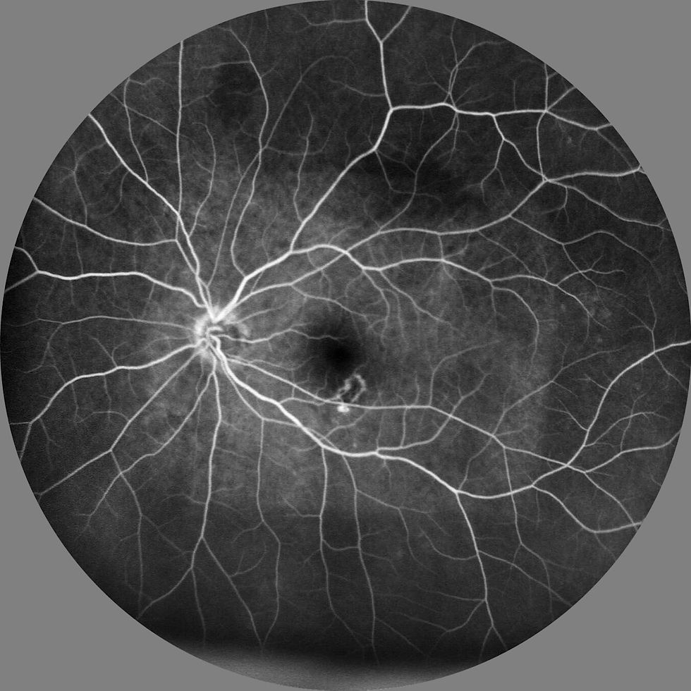
Fluorescein angiography - a special dye is injected into the bloodstream, and photographs with specific light wavelengths are taken to assess the blood vessels in the retina.
Optical coherence tomography (OCT) - a non-invasive imaging technique that captures detailed 'slices' of the retina, allowing the doctor to assess the macula and layers of the retina.
Treatment
While there is no perfect cure for AMD, several treatments can help manage and slow its progression:
Anti-VEGF injections:
Medications like ranibizumab, aflibercept, and bevacizumab are injected into the eye to block vascular endothelial growth factor (VEGF), which causes abnormal blood vessel growth that can damage the retina.
Photodynamic therapy (PDT):
A light-sensitive drug is injected into the bloodstream and activated by a laser to target and shrink abnormal blood vessels in the retina.
Laser therapy:
In some cases, high-energy laser beams are used to destroy abnormal blood vessels and prevent further damage.
Nutritional supplements:
The use of antioxidants and zinc (found in AREDS and AREDS2 formulas) can reduce the risk of progression in people with early-stage AMD.
For dry AMD, there are no direct treatments, but managing risk factors like smoking cessation, diet modifications, and monitoring for any signs of progression is key.
Prognosis
The prognosis for AMD varies depending on the type (dry or wet) and the stage at diagnosis. Dry AMD tends to progress slowly, while wet AMD can lead to rapid and severe vision loss if left untreated. Regular monitoring and timely treatment are crucial in maintaining vision and quality of life. Early intervention, especially for wet AMD, can help prevent further vision loss. However, advanced AMD can lead to permanent central vision loss, although peripheral vision is typically preserved.
Wet AMD vs Dry AMD
Dry AMD is charaterised by the gradual breakdown and thinning of macular cells and the accumulation of yellow deposits called drusen under the retina. The progression is typically slow and gradual (over years) and vision loss tends to be (but not always) mild to moderate. Studies have shown that nutritional supplements (like those based on the AREDS2 formula) may help slow progression.
In both cases, regular monitoring would coherently give better outcomes through early detection and management.
Dry AMD can sometimes progress into wet AMD.
Wet AMD accounts for the majority of severe vision loss from AMD.
Regular eye exams are critical for early detection and management.

Conclusion
AMD is a serious eye condition that can significantly impact quality of life, particularly in older adults. While there is no cure, various treatments can help manage the condition, slow progression, and preserve vision. Early detection and regular monitoring are essential for those at risk, as timely intervention can significantly improve outcomes.