
Fundus Fluorescein Angiography / Indo-Cyan Green Angiography
- Jan 13
- 3 min read
Updated: Mar 3
FFA (Fundus Fluorescein Angiography, also called Fluorescein Angiography or FA) and ICGA (Indocyanine Green Angiography, also called ICG Angiography) are specialised diagnostic imaging tests used in ophthalmology to evaluate the blood vessels in the back of the eye. They help diagnose and monitor conditions affecting the retina (the light-sensitive layer) and choroid (the vascular layer beneath the retina).
Why do we do this test?
These two tests are typically done to look at the blood circulation in the two respective layers (the retina and the choroid), which will allow the doctor to check for any leakage or blocked vessels, which can occur in certain eye conditions - such as macular oedema in diabetic retinopathy, neovascularisation (growth of new blood vessels) in wet AMD (age-related macular degeneration) or a retinal artery or branch occlusion such as a BRVO (branch retinal vein occlusion).
What is the procedure like?
Preparation - we will dilate your pupils (if they aren't already dilated, and capture preliminary images of the back of your eye. Throughout the procedure, you will have to position your head on the headrest and chinrest in front of the imaging machine.
Injection of dye and capturing of images - a small needle will be placed into your vein, commonly on the back of your hand, for a dye to be injected (either the sodium fluorescein dye for FFA or indocyanine green dye for ICGA).
Right immediately after the dye is injected, a series of images or video will begin the capture as the dye circulates through the bloodstream into the eye structures. It is important to keep looking at the target or direction as instructed. This will go on for about 15 minutes with images taken at regular intervals.
What should I expect when the dye is being injected?
It is normal to feel the cool sensation of the dye going up your arm, which may then cause the feeling of a hot flush. A small percentage of patients may experience a metallic taste or nausea. Should you feel major discomfort at any time during the procedure, you should let us know about it.
What should I expect post-procedure?
With multiple images taken, you may see after-images or feel that your vision is darker than usual. This typically lasts for a few minutes and recover is usually very quick.
If FFA dye is used, your skin may turn slightly yellowish with a gradual recovery back to normal in 1-2 days. Your urine may also be bright yellow fluorescent as the body clears the dye from the circulatory system. Stay well-hydrated.
The effects of the dilation drops, such as light sensitivity and difficulty with near work or adjusting focus, will typically wear off after a few hours. You should avoid driving and consider wearing sunglasses to help with the feeling of glare.
Are there side effects and risks?
While this is considered a generally safe procedure, there may be minor side effects such as itching and mild pain at the injection site, discomfort from dye leakage under the skin, and possible severe allergic reaction to the dye.
You should inform us if you have any shellfish or iodine allergy or kidney issues. (Some precautions may be a myth, a study shows)
Your doctor will discuss any personal risks (e.g., if you have shellfish allergies for ICG or kidney issues). These tests provide critical information to guide treatments like injections, laser therapy, or monitoring. If you're scheduled for one, ask your ophthalmologist for personalized details

I love technical information, tell me more!
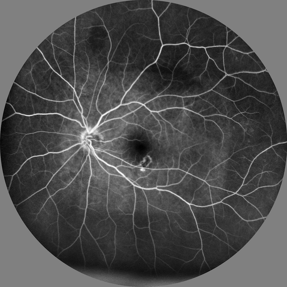
Fundus Fluorescein Angiography (FFA) is the gold-standard imaging technique for visualizing retinal blood vessels, providing high-contrast images by using blue light (465–490 nm) to excite intravenously injected sodium fluorescein dye, which emits yellow-green fluorescence (520–530 nm). It delineates vessel architecture, including arteries, capillaries, and veins, typically revealing hyperfluorescent filling patterns in early phases (arterial to arteriovenous) that progress to leakage or staining in later phases, crucial for diagnosing conditions like diabetic retinopathy, age-related macular degeneration, and retinal vein occlusions.
Indocyanine Green Angiography (ICGA) is the gold-standard imaging technique for visualizing choroidal blood vessels, providing high-contrast images by using infrared light (835 nm) to penetrate melanin and fluid. It identifies vessel architecture, including arteries, choriocapillaris, and veins, typically revealing hyperfluorescent vascular patterns in early phases that become more diffuse later, crucial for diagnosing conditions like polypoidal choroidal vasculopathy (PCV).


